Zebrafish models for innate immune research
In my career, I have developed and validated zebrafish embryonic and larval models that enable real-time tracking and quantification of macrophage behavior within a whole living disease context. These include models of mycobacterial and fungal infection, as well as orthotopic and metastatic (patient-derived) cancer xenografts. The development of these models is favored by their alignment with the 3Rs principle of animal research, promoting ethical and responsible research practices. However, it is crucial to be aware of the nuances and limitations of different experimental models, and in translational research, mechanistic insights are only valuable if the models in which they are discovered allow their therapeutic translation. This is why I have focused an important part of my career on studying and improving the translatability of these models.
In my first first-author publication, we characterized 8 complement c3 genes in zebrafish, including a teleost-specific duplication event that gave rise to an exclusive paralog with antagonistic immune function, contrasting with only one C3 gene in humans. Lack of 1-to-1 orthology is a common occurrence among immune genes in zebrafish, as we characterized for interferon-induced proteins with tetratricopeptide repeats (ifits), perforins, or caspases and inflammasome components (Figure 1). How do we translate the function of genes that do not exist in another organism? If one of the main components of the complement cascade system, sitting at the base of inflammatory processes, does not translate, what does it mean for all inflammatory research in zebrafish? This is particularly significant given the debates questioning the applicability of murine models for studying human inflammatory diseases due to controversial differences in gene modulation during inflammation. If murine models are questioned, what is the relevance of evolutionarily distant species for modeling human diseases?
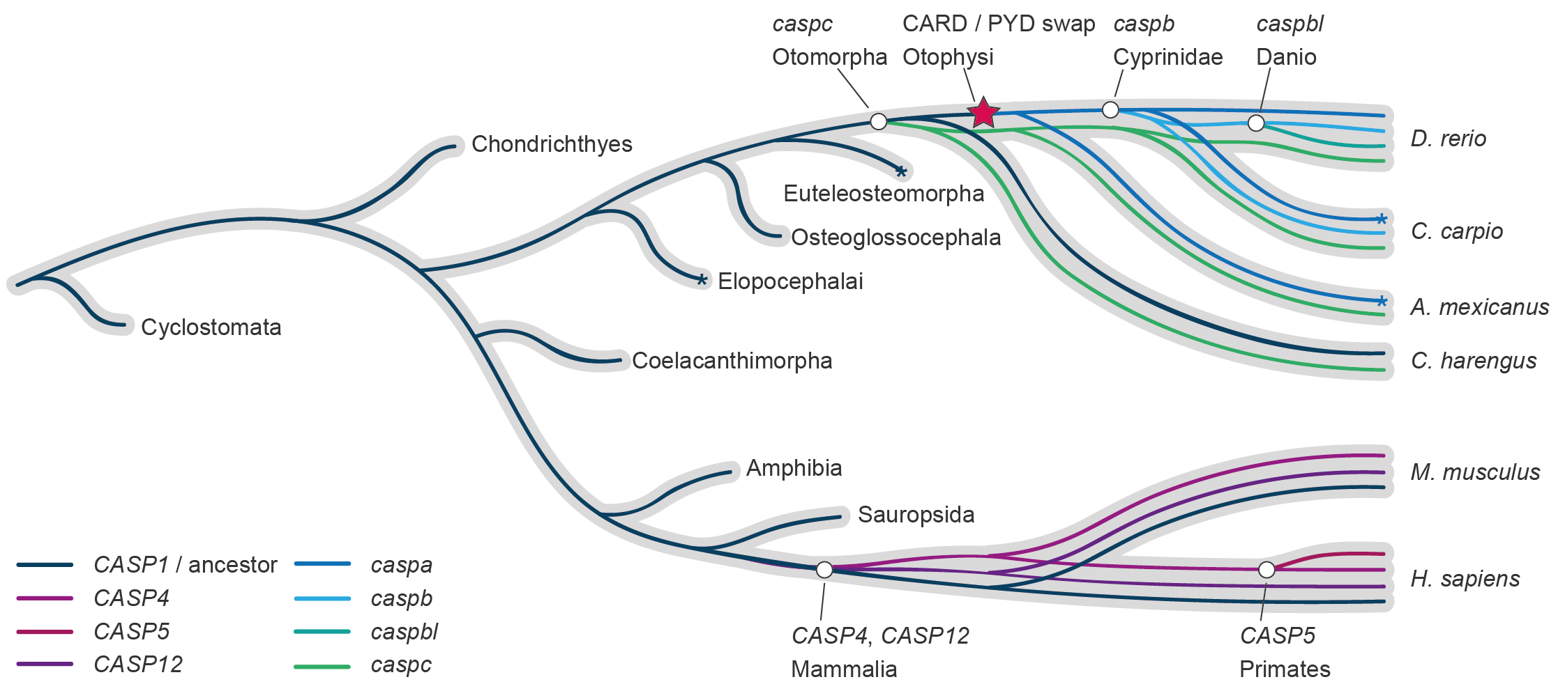
Figure 1. Reconciliation of the zebrafish and human caspase-1 gene evolution. Both the zebrafish and human caspase-1 genes have undergone independent duplication events, resulting in the presence of 4 caspase-1 family genes in each species, but with distinct evolutionary trajectories. From Forn-Cuní et al., Cells, 2019. DOI: 10.3390/cells8080901
During my PhD, I showed that, despite species-specific sensitivities and physiologies (like the c3 paralogs characterized above), zebrafish exhibit a conserved inflammatory response, mirroring key aspects of mammalian inflammatory signaling, formally validating its translational potential for inflammatory research. This lesson has guided part of my career: in my pursuit of bringing zebrafish studies into clinical relevance, my focus has broadened from specific genes (e.g., c3 duplications, perforins) to pathways (e.g., the acute inflammatory response or oxidative phosphorylation), to cellular and tissue dynamics (e.g., the innate immune response to Aspergillus conidia, macrophage recruitment and angiogenesis), while the spatiotemporal resolution has increased from whole organism (bulk RNA expression during development), to tissues (liver- or kidney-specific responses), to specific cell types inside a whole-organism context (phagocytes interacting with conidia at infection onset, single-cell transcriptomics).
Video 1. Timelapse showing macrophage-mediated (magenta) angiogenesis in a zebrafish larvae with fluorescent blood vessels (cyan) after engraftment of human glycolytic tumor cells (yellow). From Yin and Forn-Cuní et al., Angiogenesis, 2024. DOI: 10.1007/s10456-024-09930-y
While I am fully convinced that the zebrafish constitutes an exceptional platform for real-time study of the bases of immunometabolism regulation in vivo, I am equally aware that current models may not fully recapitulate the complex metabolic landscape of human macrophages and that, currently, the translation from zebrafish to the clinic is challenging. That is why I believe that, to study innate immune cell interactions, zebrafish models excel in an integrated setting with mice and human data.